STEMdiff™ Ventricular Cardiomyocyte Differentiation Kit

Serum-free media for differentiation of human PSCs to ventricular cardiomyocytes and long-term maintenance of human PSC-derived cardiomyocytes
Need a high-quality cell source? Choose from our hiPSC healthy control lines, manufactured with mTeSR™ Plus.

STEMdiff™ Ventricular Cardiomyocyte Differentiation Kit

Serum-free media for differentiation of human PSCs to ventricular cardiomyocytes and long-term maintenance of human PSC-derived cardiomyocytes

From: 814 USD
Catalog #
(Select a product)
Serum-free media for differentiation of human PSCs to ventricular cardiomyocytes and long-term maintenance of human PSC-derived cardiomyocytes
Add to Wish List

Product Advantages


  • Supports the entire hPSC-derived cardiomyocyte workflow

  • Simple monolayer protocol produces cardiomyocytes in 15 days

  • One serum-free kit generates over 50 million cardiomyocytes (cTnT+)

  • Robust performance with minimal variability across multiple hPSC lines

What's Included

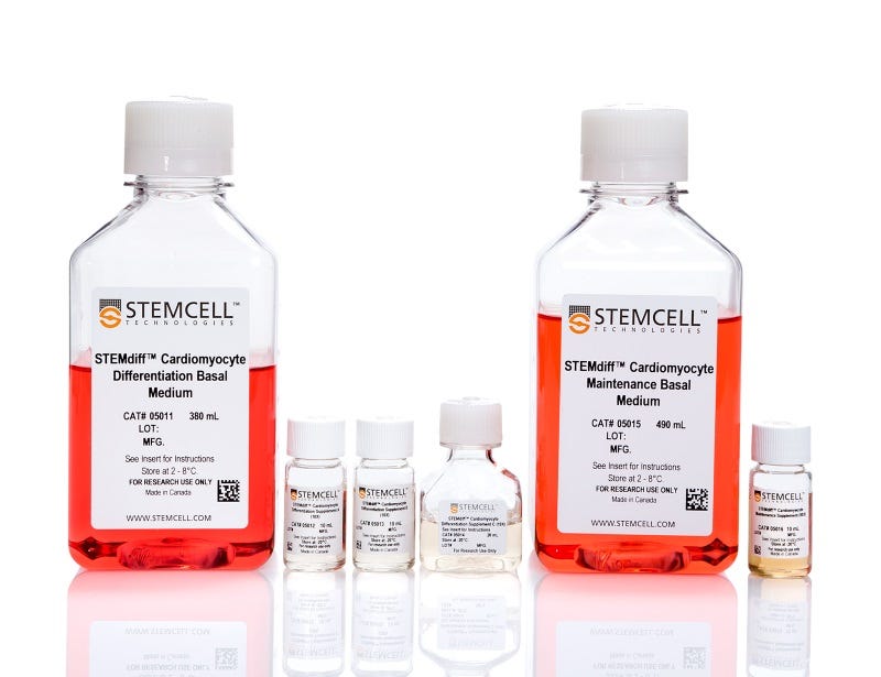
  • STEMdiff™ Cardiomyocyte Differentiation Basal Medium, 380 mL
  • STEMdiff™ Ventricular Cardiomyocyte Differentiation Supplement A (10X), 10 mL
  • STEMdiff™ Ventricular Cardiomyocyte Differentiation Supplement B (10X), 10 mL
  • STEMdiff™ Ventricular Cardiomyocyte Differentiation Supplement C (10X), 20 mL
  • STEMdiff™ Cardiomyocyte Maintenance Basal Medium, 490 mL
  • STEMdiff™ Cardiomyocyte Maintenance Supplement (50X), 10 mL
Products for Your Protocol
To see all required products for your protocol, please consult the Protocols and Documentation.

Overview

STEMdiff™ Ventricular Cardiomyocyte Differentiation Kit (Catalog #05010) includes a medium for differentiation of human embryonic stem (ES) and induced pluripotent stem (iPS) cells (human pluripotent stem cells [hPSCs]) into ventricular cardiomyocytes (cardiac troponin T-positive [cTnT+]), as well as a medium for maintenance of hPSC-derived cardiomyocytes. This serum-free kit can be used to generate ventricular cardiomyocytes derived from a clump culture of hPSCs maintained in mTeSR™1 (Catalog #85850), mTeSR™ Plus (Catalog #100-0276), TeSR™-AOF (Catalog #100-0401), or TeSR™-E8™ (Catalog #05990). Greater than 80% of these cells will be cTnT+. An average of 1 x 10^6 cells can be harvested from a single well of a 12-well plate.

STEMdiff™ Cardiomyocyte Maintenance Kit (Catalog #05020) comprises the maintenance basal medium and supplement; it can be used for long-term maintenance of hPSC-derived cardiomyocytes for one month or longer. These cardiomyocytes can be used in various downstream applications and analyses.

NOTE: This product was formerly named ‘STEMdiff™ Cardiomyocyte Differentiation Kit’; the product itself and manufacturing procedures have not changed, but the name has been updated to more accurately reflect the cell type generated.
Subtype
Specialized Media
Cell Type
Cardiomyocytes, PSC-Derived
Species
Human
Application
Cell Culture, Differentiation, Maintenance
Brand
STEMdiff
Area of Interest
Disease Modeling, Drug Discovery and Toxicity Testing, Stem Cell Biology
Formulation Category
Serum-Free

Data Figures

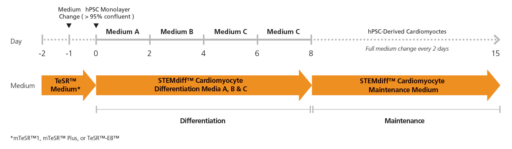
Figure 1. Cardiomyocyte Differentiation Protocol

Two days before the differentiation protocol, hPSC colonies are harvested and seeded as single cells at 350,000 cells/well in a 12-well format in TeSR™ medium. After one day (Day -1), the medium is replaced with fresh TeSR™ medium. The following day (Day 0), the TeSR™ medium is replaced with Medium A (STEMdiff™ Cardiomyocyte Differentiation Basal Medium containing Supplement A) to begin inducing the cells toward a cardiomyocyte fate. On day 2, a full medium change is performed with fresh Medium B (STEMdiff™ Cardiomyocyte Differentiation Basal Medium containing Supplement B). On days 4 and 6, full medium changes are performed with fresh Medium C (STEMdiff™ Cardiomyocyte Differentiation Basal Medium containing Supplement C). On day 8, medium is switched to STEMdiff™ Cardiomyocyte Maintenance Medium with full medium changes on days 10, 12 and 14, to promote further differentiation into cardiomyocyte cells. Small beating areas of cardiomyocytes can be seen as early as day 8, progressing to a full lawn of beating cardiomyocytes that can be harvested as early as day 15.

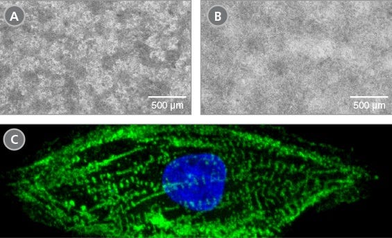
Figure 2. Morphology of hPSC-Derived Cardiomyocytes

Representative images of (A) hES (H9) cells and (B) hiPS (WLS-1C) cells on day 15 of differentiation to cardiomyocytes using the STEMdiff™ Ventricular Cardiomyocyte Differentiation Kit. Differentiated cells exhibit typical cardiomyocyte morphology as an adherent, tightly packed web-like monolayer of beating cells. (C) Representative confocal microscopy image of a single hPSC-derived cardiomyocyte generated with the STEMdiff™ Ventricular Cardiomyocyte Differentiation Kit and stained with cTnT (green) and DAPI (blue).

Figure 3. Efficient and Robust Generation of cTnT-Positive Cardiomyocytes

hES and hiPS cells were cultured for 15 days in single wells of 12-well plates using the STEMdiff™ Ventricular Cardiomyocyte Differentiation Kit. At the end of the culture period, cells were harvested and analyzed by flow cytometry for expression of cardiac troponin T (cTnT). (A) Histogram analysis for cardiomyocyte cell marker cTnT for cultures of hES (H9) and hiPS (WLS-1C and STiPS-M001) cells. (Filled = sample; blank = secondary antibody only control) (B,C) Percentages and total numbers of cells expressing cTnT in cultures of hES or hiPS cells are shown. Data shown as mean ± SEM; n=3.

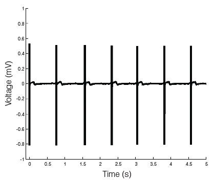
Figure 4. hPSC-Derived Cardiomyocytes Exhibit a Robust and Stable Excitability Profile

Microelectrode array (MEA) voltage recordings of cardiomyocytes (day 27) derived from human pluripotent stem cells generated and maintained with the STEMdiff™ Cardiomyocyte Differentiation and Maintenance Kits. The hPSC-derived cardiomyocytes have a characteristic electrical profile and stable beat rate. A large depolarization spike followed by a smaller repolarization deflection is observed.

Microelectrode array and flow cytometry of human ES and iPS cells maintained in mTeSR™1 (daily feeds) or mTeSR™ Plus (restricted feeds) and differentiated to cardiomyocytes using the STEMdiff™ Ventricular Cardiomyocyte Differentiation Kit.

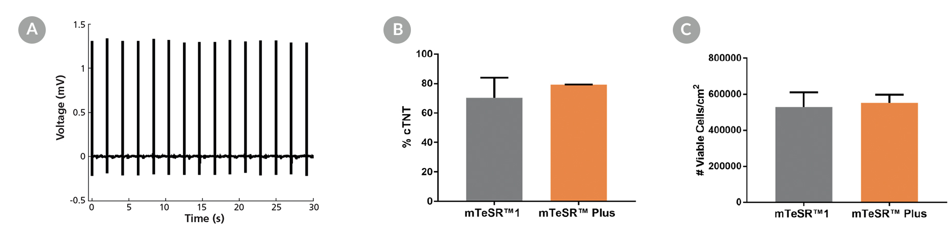
Figure 5. Generation of Cardiomyocytes from hPSCs Maintained in mTeSR™ Plus

Human ES (H9) and iPS (WLS-1C) cells were maintained in mTeSR™1 (daily feeds) or mTeSR™ Plus (restricted feeds) and differentiated to cardiomyocytes using the STEMdiff™ Ventricular Cardiomyocyte Differentiation Kit. At the end of the differentiation period, cells were harvested and analyzed by microelectrode array (MEA) and flow cytometry. (A) Representative MEA voltage recordings of cardiomyocytes (day 20) demonstrate a characteristic electrical profile and stable beat rate. (B) Percentages of cells expressing cTNT and (C) total number of viable cells harvested are shown. Data are expressed as the mean (± SEM); n=2.

Microscopy images of iPSCdirect cells and differentiated ventral cardiomyocytes, and a video of coordinated contraction or beating behavior of cardiomyocytes in a culture dish

Figure 6. iPSCdirect™ SCTi003-A Human Pluripotent Stem Cells Can Successfully Differentiate into Ventricular Cardiomyocytes

Ventricular cardiomyocytes were generated from iPSCdirect™ SCTi003-A cells using STEMdiff™ Ventricular Cardiomyocyte Differentiation Kit (Catalog #05010). (A) 48 hours after thawing and plating in mTeSR™ Plus and CloneR™2, iPSCdirect™ cells reached the desired confluency and are ready for Day 0 of differentiation according to the STEMdiff™ Ventricular Cardiomyocyte Product Information Sheet. (B) By Day 15 of differentiation, monolayer cultures show iPSC-derived ventricular cardiomyocytes that (C) exhibit coordinated beating behavior.

Protocols and Documentation

Find supporting information and directions for use in the Product Information Sheet or explore additional protocols below.

Document Type
Product Name
Catalog #
Lot #
Language
Catalog #
05010
Lot #
All
Language
English
Document Type
Safety Data Sheet 1
Catalog #
05010
Lot #
All
Language
English
Document Type
Safety Data Sheet 2
Catalog #
05010
Lot #
All
Language
English
Document Type
Safety Data Sheet 3
Catalog #
05010
Lot #
All
Language
English
Document Type
Safety Data Sheet 4
Catalog #
05010
Lot #
All
Language
English
Document Type
Safety Data Sheet 5
Catalog #
05010
Lot #
All
Language
English
Document Type
Safety Data Sheet 6
Catalog #
05010
Lot #
All
Language
English

Applications

This product is designed for use in the following research area(s) as part of the highlighted workflow stage(s). Explore these workflows to learn more about the other products we offer to support each research area.

Resources and Publications

Educational Materials (17)

Publications (24)

ALY688 Protects Against Myocardial Ischemia-Reperfusion Injury via Direct Effects and Rab8a-Dependent Extracellular Vesicles. H. Sung et al. Journal of extracellular vesicles 2026 Sep

Abstract

Despite advances in percutaneous coronary intervention, ischemia-reperfusion (IR) injury remains a major cause of morbidity and mortality. Adiponectin confers broad cardioprotective effects, motivating the development of adiponectin receptor agonists. Here, we investigated the cardioprotective efficacy and mechanisms of ALY688, a synthetic adiponectin receptor agonist peptide, in myocardial IR injury. In a clinically translatable rat IR model, intravenous administration of ALY688 during ischemia together with subcutaneous dosing that continued for 28 days reduced troponin-I levels, cardiomyocyte death, and infarct size, while preserving cardiac function. ALY688 restored autophagic flux, mitigated reactive oxygen species accumulation, and suppressed apoptosis in both IR hearts and hypoxia-reoxygenation (HR)-treated cardiomyocytes. Proteomic profiling revealed that Rab8a, downregulated by IR, was maintained with ALY688 treatment. Notably, ALY688 increased extracellular vesicle (EV) abundance in myocardium and plasma, and EVs from treated animals displayed distinct proteomic signatures enriched in glycolytic and oxidative stress-related proteins. These EVs conferred protection against HR-induced injury in H9c2 and human iPSC-derived cardiomyocytes. CRISPR-mediated Rab8a knockout impaired ALY688-induced EV biogenesis and attenuated the cytoprotective effects of these EVs. Collectively, these findings identify ALY688 as a promising therapeutic that mitigates IR injury via both direct myocardial protection and Rab8a-dependent EV-mediated cardioprotective signalling.
Dose-dependent sensitivity of human three-dimensional chromatin to a heart disease-linked transcription factor. Z. Grant et al. Science (New York, N.Y.) 2026 Jul

Abstract

Dosage-sensitive transcription factors (TFs) underlie altered gene regulation in human developmental disorders, and cell type-specific gene regulation is linked to the reorganization of three-dimensional (3D) chromatin during cellular differentiation. In this work, we show dose-dependent regulation of chromatin organization by the congenital heart disease (CHD)-linked, lineage-restricted TF TBX5 in human cardiomyocyte differentiation. Genome organization, including compartments, topologically associated domains, and chromatin loops, was sensitive to reduced TBX5 dosage in a human model of CHD, with variations in response across individual cells. Cohesin binding was reduced at TBX5-bound enhancer elements in a TBX5 dose-dependent manner, providing a potential mechanism for disrupted loop formation. These results highlight the importance of lineage-restricted TF dosage in cell type-specific 3D chromatin dynamics, suggesting a mechanism for TF-dependent disease.
AAV Vector Toolkit for the Delivery and Expression of the Artificial microRNA in the Murine Heart. I. Galkin et al. Biotech (Basel (Switzerland)) 2026 Jul

Abstract

BACKGROUND: Adeno-associated virus (AAV) vector-mediated gene transfer is an emerging treatment strategy for severe cardiac disorders with genetic etiology. We refined the AAV toolkit to achieve efficient and selective expression of the therapeutic transgene in mouse hearts. METHODS: Using vectors with a reporter transgene, we evaluated AAV administration routes, AAV serotype tropism to the myocardium, and cardiospecific promoters. RESULTS: We showed that systemic AAV administration provides potent delivery and uniform transduction of cardiac tissue, outperforming localized injection techniques. The MyoAAV 2A capsid variant enabled an improved heart-to-liver transduction ratio compared to the parental AAV9 serotype. Screening a panel of cardiac and pan-muscular promoters in vitro and in vivo verified the superiority of the cardiac troponin T (cTnT) promoter for robust heart-specific transgene expression. Finally, we demonstrated that the cumulative properties of systemic AAV delivery, the MyoAAV 2A serotype, and the cTnT promoter allowed for efficient cardiac synthesis of the therapeutic transgene-an artificial miRNA designed for the gene suppression strategy of FLNC-related cardiomyopathy. CONCLUSIONS: Our findings establish an effective AAV approach for transgene transfer into the mouse heart and promote the development of gene therapy for cardiac disorders.
Need a high-quality cell source? Choose from our hiPSC healthy control lines, manufactured with mTeSR™ Plus.