PneumaCult™-ALI Medium

Serum- and BPE-free medium for human airway epithelial cells cultured at the air-liquid interface
Need robust HAECs for differentiation? Begin your workflow by expanding HAECs in PneumaCult™-NGEx Medium for optimal ALI cultures.

PneumaCult™-ALI Medium

Serum- and BPE-free medium for human airway epithelial cells cultured at the air-liquid interface

From: 306 USD
Catalog #
(Select a product)
Serum- and BPE-free medium for human airway epithelial cells cultured at the air-liquid interface
Add to Wish List

Product Advantages


  • HBECs cultured with PneumaCult™-ALI undergo extensive mucociliary differentiation to form a pseudostratified epithelium that closely resembles the human airway

  • PneumaCult™-ALI is serum-free and BPE-free to minimize variability

What's Included

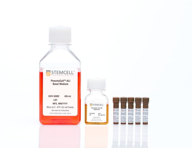
  • PneumaCult™-ALI Basal Medium, 450 mL
  • PneumaCult™-ALI 10X Supplement, 50 mL
  • PneumaCult™-ALI Maintenance Supplement (100X), 5 x 1 mL
Products for Your Protocol
To see all required products for your protocol, please consult the Protocols and Documentation.

Overview

PneumaCult™-ALI Medium (Catalog #05001) is a serum- and BPE-free medium for the culture of human airway epithelial cells at the air-liquid interface (ALI). Airway epithelial cells cultured in PneumaCult™-ALI Medium undergo extensive mucociliary differentiation to form a pseudostratified epithelium that exhibits morphological and functional characteristics similar to those of the human airway in vivo. PneumaCult™-ALI Medium is also available in a kit that includes 12 mm Transwell® inserts (Catalog #05021) or 6.5 mm Transwell® inserts (Catalog #05022).

Together, PneumaCult™-ALI Medium and PneumaCult™-NGEx Medium (Catalog #100-1505) constitute a fully integrated BPE-free culture system for in vitro human airway modeling that is also compatible with primary human nasal epithelial cells. This robust and defined system is a valuable tool for basic respiratory research, toxicity studies, and drug development.

Learn how to culture human airway epithelial cells at the ALI in our On-Demand Pulmonary Course or browse our Frequently Asked Questions (FAQs) about the ALI culture workflow using PneumaCult™.

For information about introductory offers to try PneumaCult™ in your lab, fill out this form.
Subtype
Specialized Media
Cell Type
Airway Cells
Species
Human
Application
Cell Culture, Differentiation, Maintenance, Organoid Culture
Brand
PneumaCult
Area of Interest
Disease Modeling, Drug Discovery and Toxicity Testing, Epithelial Cell Biology
Formulation Category
Serum-Free

Data Figures

Figure 1. Overview of the PneumaCult™ Culture System

Expansion of human bronchial epithelial cells (HBECs) in submerged culture is performed with PneumaCult™-Ex Plus or PneumaCult™-Ex. During the early Expansion Phase of the ALI culture procedure, PneumaCult™-Ex Plus or PneumaCult™-Ex is applied to the apical and basal chambers. Upon reaching confluence, the culture is air-lifted by removing the culture medium from both chambers, and adding PneumaCult™-ALI to the basal chamber only. Differentiation into a pseudostratified mucociliary epithelium is obtained following 21-28 days of incubation and can be maintained for more than one year.

Figure 2. HBECs Cultured in PneumaCult™-Ex Successfully Differentiate into a Pseudostratified Mucociliary Epithelium with PneumaCult™-ALI

Early-passage (P1-3) HBECs cultured in PneumaCult™-Ex successfully differentiate when cultured at air-liquid interface with PneumaCult™-ALI for 28 days. H&E staining revealed the pseudostratifi ed structure of the epithelium with cilia present at the apical surface (A). Periodic acid-Schiff staining demonstrated the presence of goblet cells (B). The presence of ciliated and goblet cells was also demonstrated by immunofl uorescence staining of cilia marker acetylated (AC)-Tubulin (green; C) and the goblet cell marker Mucin5AC (green; D). Appropriate positioning of basal cells along the transwell insert was visualized by immunofl uorescence staining using the basal cell markers p75NTR (green) and p63 (red; E,F). A representative merged image indicates the apical cells, detected by DAPI alone, positioned along the epithelium and in close contact with the basal cells (detected by DAPI, p63 and p75NTR co-labeling) located along the insert (G).

Figure 3. Electrophysiological characterization of differentiated HBECs (P4) that were expanded in PneumaCult™-Ex Plus, PneumaCult™-Ex, and Bronchial Epithelial Growth Media

TEER (A) and representative characterization of the ion channel activities (B) for ALI cultures at 28 days post air-lift using HBECs expanded in PneumaCult™-Ex Plus, PneumaCult™-Ex, or Bronchial Epithelial Growth Media. Amiloride: ENaC inhibitor. IBMX and Forskolin: CFTR activators. Genistein: CFTR potentiator. CFTRinh-172: CFTR inhibitor. UTP: Calciumactivated Chloride channels (CaCCs) activator. All ALI differentiation cultures were performed using PneumaCult™-ALI.

Protocols and Documentation

Find supporting information and directions for use in the Product Information Sheet or explore additional protocols below.

Document Type
Product Name
Catalog #
Lot #
Language
Catalog #
05001
Lot #
All
Language
English
Catalog #
05021
Lot #
All
Language
English
Catalog #
05022
Lot #
All
Language
English
Document Type
Safety Data Sheet 1
Catalog #
05001
Lot #
All
Language
English
Document Type
Safety Data Sheet 2
Catalog #
05001
Lot #
All
Language
English
Document Type
Safety Data Sheet 3
Catalog #
05001
Lot #
All
Language
English
Document Type
Safety Data Sheet 1
Catalog #
05021
Lot #
All
Language
English
Document Type
Safety Data Sheet 2
Catalog #
05021
Lot #
All
Language
English
Document Type
Safety Data Sheet 3
Catalog #
05021
Lot #
All
Language
English
Document Type
Safety Data Sheet 1
Catalog #
05022
Lot #
All
Language
English
Document Type
Safety Data Sheet 2
Catalog #
05022
Lot #
All
Language
English
Document Type
Safety Data Sheet 3
Catalog #
05022
Lot #
All
Language
English

Applications

This product is designed for use in the following research area(s) as part of the highlighted workflow stage(s). Explore these workflows to learn more about the other products we offer to support each research area.

Resources and Publications

Educational Materials (27)

On-Demand Training

Publications (155)

Blocking AGEs reduces airway epithelial viral susceptibility in diabetes-exacerbated COPD. Z. Chen et al. EMBO molecular medicine 2026 Sep

Abstract

Although diabetic complications are well-established in multiple organs, their impact on the respiratory system remains unclear. Clinical evidence indicates accelerated chronic obstructive pulmonary disease (COPD) progression in individuals with diabetes, yet the underlying mechanisms are poorly defined. Using single-cell RNA sequencing of airway epithelium, we identified distinct transcriptional alterations in COPD patients with or without diabetes, particularly in host-defense pathways related to viral infection, which is a key driver of COPD exacerbations. Functionally, hyperglycemia enhanced influenza virus susceptibility in both COPD-derived epithelial cells and COPD mice with diabetes, indicating compromised airway defense. We further demonstrated that hyperglycemia promotes advanced glycation end products (AGEs) accumulation in airway epithelium, with AGEs levels correlating with infection severity. Pharmacological inhibition of AGEs formation with aminoguanidine significantly reduced viral load both in vitro and in vivo. Mechanistically, AGEs activate p38/ERK/IKK proinflammatory signaling via RAGE and MD2 receptors, leading to epithelial injury and heightened viral susceptibility. Our findings reveal a hyperglycemia-AGEs-RAGE/MD2 axis that increases viral susceptibility in COPD, highlighting AGEs targeting as a promising therapeutic strategy for COPD patients with diabetes.
CRSwNP-derived cells retain native disease-relevant characteristics in vitro P. Kühnel et al. Journal of Inflammation (London, England) 2026 Mar

Abstract

Objective and designChronic rhinosinusitis (CRS) is a heterogeneous inflammatory disease of the paranasal sinuses, which is divided into CRS with nasal polyps (CRSwNP) and CRS without nasal polyps (CRSsNP). CRSwNP is typically caused by type 2 inflammation, which is characterized by elevated IL-4 and IL-13 levels, impairment of the epithelial barrier, and tissue remodeling. While the involvement of immune cells is well known, it remains unclear to what extent structural cells intrinsically maintain disease-specific functional programs. The aim of this study was to determine whether epithelial cells and fibroblasts derived from CRSwNP and CRSsNP differ in their barrier properties, inflammatory reactivity, and type 2-associated functional characteristics.MethodsAir–liquid interface (ALI) epithelial cultures and primary fibroblast cultures were generated from CRSwNP and CRSsNP tissue. Epithelial barrier integrity was assessed by transepithelial electrical resistance (TEER), and inflammatory responses to TLR stimulation were analyzed by qRT-PCR. Fibroblast migration was evaluated using scratch assays. Cellular responses to IL-4/IL-13 with or without Dupilumab were quantified by qRT-PCR.ResultsCRSwNP-derived epithelial cells exhibited delayed tight junction formation and impaired differentiation compared to CRSsNP cells. Poly(I:C) stimulation induced stronger expression of Th2-associated cytokines in CRSwNP cultures. CRSwNP fibroblasts showed reduced migratory capacity and a heightened induction of Th2 cytokines and extracellular matrix genes following IL-4/IL-13 stimulation relative to CRSsNP fibroblasts.ConclusionEpithelial cells and fibroblasts derived from CRSwNP retain disease-associated type 2 characteristics in vitro, indicating persistent disease-aligned programmed functional alterations of the polyp microenvironment. In contrast, CRSsNP-derived cells lacked comparable enhanced type 2 responsiveness. These findings support CRSwNP as a distinct, self-sustaining inflammatory endotype and underscore the value of patient-derived models for investigating disease mechanisms and targeted therapies.Supplementary InformationThe online version contains supplementary material available at 10.1186/s12950-026-00497-7.
Neutrophil myeloperoxidase as a functional biomarker for RSV severity: implications for in vitro therapeutic screening. M. Palor et al. Nature communications 2026 Jul

Abstract

Respiratory syncytial virus (RSV) is a leading cause of severe lower respiratory tract infections in infants, yet therapeutics are lacking. The aim of this study was to develop a pre‑clinical model that recapitulates key clinical outcomes in infants with RSV bronchiolitis, such as neutrophil activation and migration into the airways. Peripheral blood neutrophils from infants with severe RSV disease admitted to the Paediatric Intensive Care Unit showed elevated myeloperoxidase (MPO) in children with RSV, compared to age-matched controls. To mechanistically model this response, we established an air-liquid interface (ALI) system incorporating paediatric airway epithelial cells, endothelial cells and neutrophils from adults, to recapitulate the blood-airway barrier. Following RSV infection, with and without treatment with antivirals remdesivir or RSV604, neutrophil migration and activation were assessed using flow cytometry. While both drugs reduced viral load, only RSV604 attenuated MPO expression. This model suggests that MPO could be useful as a readout of therapeutic efficacy. Targeting neutrophil-driven inflammatory pathways may be critical for reducing pathology in infant RSV infection.
Need robust HAECs for differentiation? Begin your workflow by expanding HAECs in PneumaCult™-NGEx Medium for optimal ALI cultures.