SepMate™-50 (IVD)

Tube for density gradient centrifugation for in vitro diagnostic (IVD) applications
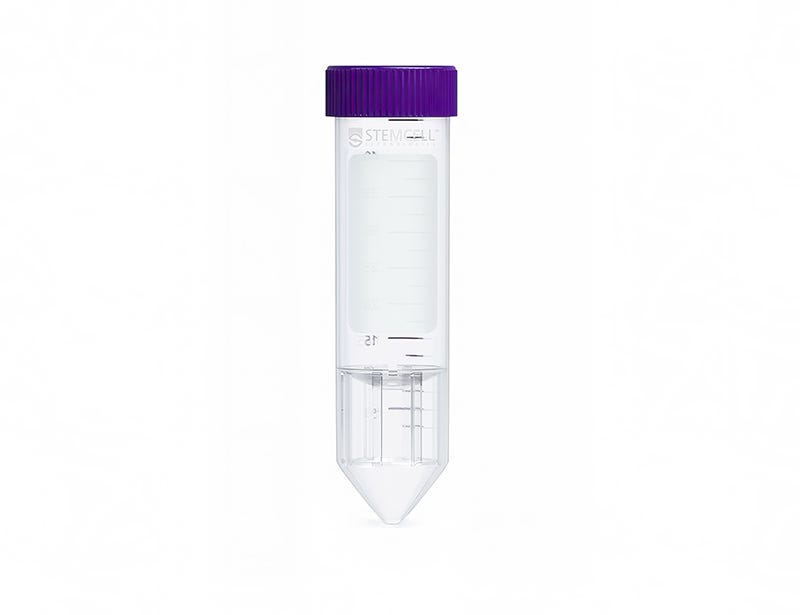
Try SepMate™-50 (IVD) tubes for density gradient centrifugation in your IVD applications. Request a Sample

SepMate™-50 (IVD)

Tube for density gradient centrifugation for in vitro diagnostic (IVD) applications

From: 670 USD
Catalog #
(Select a product)
Tube for density gradient centrifugation for in vitro diagnostic (IVD) applications
Add to Wish List

Product Advantages


  • Eliminates the need for carefully layering blood over the density gradient medium (e.g. Lymphoprep™, etc.)

  • Reduces total centrifuge time to 10 minutes with the brake on for fresh samples

  • Allows fast and easy harvesting of the isolated mononuclear cells by simply pouring off the supernatant

  • Can be combined with RosetteSep™ enrichment cocktails to isolate specific cell types in just 30 minutes

What's Included

  • SepMate™-50 (IVD), 100 Tubes (Catalog #85450)
    • Dispenser box containing 4 bags, 25 Tubes/Bag
  • SepMate™-50 (IVD), 500 Tubes (Catalog #85460)
    • Dispenser box containing 4 bags, 25 Tubes/Bag (Catalog #85450) x 5

What Our Scientist Says

Traditional isolation of PBMCs requires careful layering of blood onto density gradient media prior to centrifugation. We developed SepMate™ to simplify this process, so anyone can isolate PBMCs with a simple pour while maintaining consistency across samples.

Peter MorinTechnical Scientist
Peter Morin, Technical Scientist

Overview

Simplify peripheral blood mononuclear cell (PBMC) isolation by incorporating SepMate™ into your density gradient centrifugation step.

SepMate™ tubes contain an insert that creates a barrier between the density gradient medium and blood, thus eliminating the need for careful blood layering and allowing mononuclear cells to be easily harvested with a simple pour. This product can be used with RosetteSep™ to isolate specific immune cell subsets.

SepMate™-50 is designed for processing 4 to 17 mL of sample.

SepMate™ is manufactured under cGMP and is available as an in vitro diagnostic (IVD) device in Australia, Canada, the European Union (EU), South Korea, Switzerland, Turkey, the United Kingdom (UK), and the United States. In China, SepMate™ is considered general laboratory equipment by the National Medical Products Administration (NMPA). The end user is responsible for determining whether the product is suitable for their specific application.

Browse our Frequently Asked Questions (FAQs) on SepMate™.
Contains
Polypropylene tube containing an insert
Subtype
Centrifugation Tubes
Cell Type
B Cells, Dendritic Cells, Monocytes, Mononuclear Cells, NK Cells, T Cells, T Cells, CD4+, T Cells, CD8+, T Cells, Other Subsets, T Cells, Regulatory
Species
Human
Sample Source
Bone Marrow, Whole Blood
Selection Method
Negative
Application
Cell Isolation, In Vitro Diagnostic
Brand
SepMate
Area of Interest
Chimerism, HLA, Immunology

Data Figures

PBMC recovery from fresh whole blood using SepMate™-50 versus standard density gradient centrifugation. Graph also shows PBMC recovery from a 48 hour-old sample using SepMate™. n in each group = 7

Figure 1. Recovery of mononuclear cells (MNCs) from peripheral blood using SepMate™-50 versus standard density gradient centrifiguation. Recovery of MNCs from fresh and 48-hour post blood draw enriched by density gradient centrifugation with SepMate™ (purple) or without (grey). There was no significant difference in the recovery of MNCS with and without SepMate™.

PBMC recovery from fresh whole blood using SepMate™-50 versus standard density gradient centrifugation. Graph also shows PBMC recovery from a 48 hour-old sample using SepMate™. n in each group = 7

Figure 2. Human CD4+ T Cell Isolation using SepMate™-50 and RosetteSep™ Human CD4+ T Cell Enrichment Cocktail

Protocols and Documentation

Find supporting information and directions for use in the Product Information Sheet or explore additional protocols below.

Document Type
Product Name
Catalog #
Lot #
Language
Catalog #
85460, 85450
Lot #
All
Language
MULTI

Applications

This product is designed for use in the following research area(s) as part of the highlighted workflow stage(s). Explore these workflows to learn more about the other products we offer to support each research area.

Resources and Publications

Publications (95)

Targeting TGFBR1 with the Investigational Inhibitor TP-6379 Improves Hematopoiesis in Low-Risk MDS Ex Vivo. T. Razabdouski et al. Cells 2026 Sep

Abstract

Myelodysplastic syndrome (MDS) is characterized by S100A9-mediated constitutive innate immune activation that leads to the inflammatory death of hematopoietic stem/progenitor cells (HSPCs). This suppressive microenvironment includes the secretion of TGFβ and is phenocopied by the S100A9-transgenic (Tg) mice. However, the role of the different ALK/TGFB receptors in hematopoiesis or in S100A9-mediated inflammation is not understood. Primary low-risk MDS bone marrow mononuclear cells (BM-MNCs) were analyzed in vitro for hematopoietic restoration by an ALK4/ALK5 inhibitor (Vactosertib), an ALK2 inhibitor (TP-0184/Itacnosertib) and an ALK5/TGFBR1-specific inhibitor (TP-6379). Specimens significantly responded to TP-6379 treatment restoring hematopoiesis, with a positive bias for specimens harboring spliceosomal mutations. We previously shown that S100A9 drives phenotypic pathology of MDS, and it has been known to induce and cooperate with TGFβ in this disease. Hence, we treated healthy BM-MNC with recombinant human (rh) S100A9 where TP-6379 rescued the S100A9-reduced hematopoiesis. The effect of TP-6379 was validated in vivo in S100A9-Tg mice. Similar results were obtained with K562 cells, with or without the knock-in (KI) mutation in SF3B1, with strong sensitivity to rhS100A9 that was overcome by TP-6379. Hence, directly targeting TGFBR1/ALK5 with TP-6379 restores healthy hematopoiesis in low-risk MDS preclinical models.
LPS stimulation reveals male and female dimorphism in human iPSC-derived macrophages. P. Graney et al. iScience 2026 Oct

Abstract

Clinical disparities in the inflammatory responses between males and females have been reported for decades, revealing sex-biased disease severity and patient outcomes. To better understand the intrinsic biological sex differences contributing to these disparities, we characterized the functional and transcriptomic responses of induced pluripotent stem cell (iPSC)-derived macrophages from male and female donors under control and inflammatory conditions using the bacterial antigen lipopolysaccharide (LPS). Bulk RNA sequencing identified 67 genes differentially expressed between male and female macrophages in the absence of inflammation, the majority of which mapped to the sex chromosomes. This sexual dimorphism was amplified by LPS stimulation, with genes predominantly located on autosomes, leading to divergent patterns in secreted protein production that were consistent with clinical findings following LPS exposure. Although future validation with a larger patient cohort is needed, these data collectively suggest that iPSC-derived macrophages can serve as a meaningful tool for understanding and improving treatment of sex-biased pathophysiological conditions.
Phenotypically similar but functionally distinct NK cell populations within the human maternal-fetal interface M. Frutoso et al. ImmunoHorizons 2026 Mar

Abstract

AbstractNatural killer (NK) cell function within tissues extends beyond exerting cytotoxicity, encompassing a range of functions that are just starting to become fully elucidated. In the context of human placentation, NK cells play a key role in enabling initial placentation, which is associated with the acquisition of tolerance-like properties. If and to which extent NK cells maintain these tolerance-like properties over the course of human pregnancy is still poorly understood. We asked if NK cells isolated from the decidual-placental interface of full-term human pregnancies are able to exert effector function. We observed a significant and striking lack in the ability of NK cells isolated from the decidual-placental interface (DPI) to produce interferon-g (IFN-γ) in response to the activating cytokines interleukin (IL)-12, IL-15, and IL-18. In contrast, NK cells from the decidua retained their responsiveness to cytokine-mediated activation. Notably, CD103+CD69+ tissue-resident NK cells were present in both DPI and decidua, yet exhibited distinct effector function from one another. Using high-parameter flow cytometry and single-cell sequencing, we found that this functional discrepancy was not directly predictable based on their cell surface phenotype or cell transcript. Together, our findings reveal the presence of distinct functional resident NK cell populations in 2 anatomically adjacent tissues at healthy full-term pregnancies.
Try SepMate™-50 (IVD) tubes for density gradient centrifugation in your IVD applications. Request a Sample