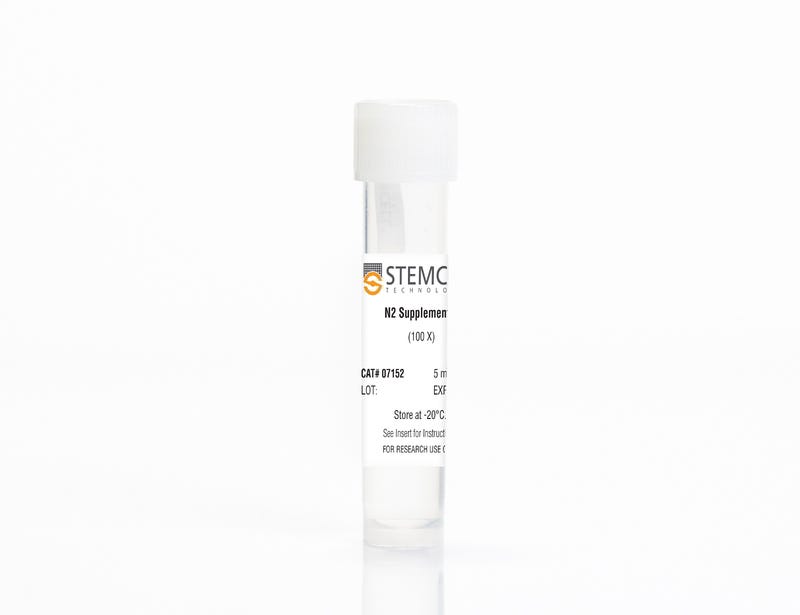
N2 Supplement-A

For neural and pancreatic differentiation of mouse and human ES and iPS cells

N2 Supplement-A

For neural and pancreatic differentiation of mouse and human ES and iPS cells

From: 113 USD
Catalog #
(Select a product)
For neural and pancreatic differentiation of mouse and human ES and iPS cells
Add to Wish List

Overview

N2 Supplement-A, containing iron-rich human transferrin, was developed for the in vitro differentiation of mouse or human embryonic stem (ES) cells and induced pluripotent stem (iPS) cells to neural and pancreatic-like cell types. Different neuronal subtypes can be generated when human ES/iPS cell-derived neural progenitor cells are cultured in BrainPhys™ Neuronal Medium (Catalog #05790) supplemented with N2 Supplement-A, NeuroCult™ SM1 Neuronal Supplement (Catalog #05711), and other factors. N2 Supplement-A is provided as a 100X stock solution.

N2 Supplement-A is available for individual sale or as a component of the BrainPhys™ Neuronal Medium N2-A & SM1 Kit (Catalog #05793).
Contains
• Recombinant human insulin
• Human holo-transferrin (iron-saturated)
• Sodium selenite
• Putrescine
• Progesterone
• Other ingredients
Subtype
Supplements
Cell Type
Endoderm, PSC-Derived, Neural Cells, PSC-Derived, Neural Stem and Progenitor Cells, Pancreatic Cells, Pluripotent Stem Cells
Species
Human, Mouse
Application
Cell Culture, Differentiation
Area of Interest
Disease Modeling, Neuroscience, Stem Cell Biology

Protocols and Documentation

Find supporting information and directions for use in the Product Information Sheet or explore additional protocols below.

Document Type
Product Name
Catalog #
Lot #
Language
Product Name
N2 Supplement-A
Catalog #
07152
Lot #
All
Language
English
Document Type
Safety Data Sheet
Product Name
N2 Supplement-A
Catalog #
07152
Lot #
All
Language
English

Applications

This product is designed for use in the following research area(s) as part of the highlighted workflow stage(s). Explore these workflows to learn more about the other products we offer to support each research area.

Resources and Publications

Publications (7)

Modelling Lyssavirus Infections in Human Stem Cell-Derived Neural Cultures. V. Sundaramoorthy et al. Viruses 2020 mar

Abstract

Rabies is a zoonotic neurological infection caused by lyssavirus that continues to result in devastating loss of human life. Many aspects of rabies pathogenesis in human neurons are not well understood. Lack of appropriate ex-vivo models for studying rabies infection in human neurons has contributed to this knowledge gap. In this study, we utilize advances in stem cell technology to characterize rabies infection in human stem cell-derived neurons. We show key cellular features of rabies infection in our human neural cultures, including upregulation of inflammatory chemokines, lack of neuronal apoptosis, and axonal transmission of viruses in neuronal networks. In addition, we highlight specific differences in cellular pathogenesis between laboratory-adapted and field strain lyssavirus. This study therefore defines the first stem cell-derived ex-vivo model system to study rabies pathogenesis in human neurons. This new model system demonstrates the potential for enabling an increased understanding of molecular mechanisms in human rabies, which could lead to improved control methods.
Maturation of Human Pluripotent Stem Cell-Derived Cerebellar Neurons in the Absence of Co-culture. T. P. Silva et al. Frontiers in bioengineering and biotechnology 2020

Abstract

The cerebellum plays a critical role in all vertebrates, and many neurological disorders are associated with cerebellum dysfunction. A major limitation in cerebellar research has been the lack of adequate disease models. As an alternative to animal models, cerebellar neurons differentiated from pluripotent stem cells have been used. However, previous studies only produced limited amounts of Purkinje cells. Moreover, in vitro generation of Purkinje cells required co-culture systems, which may introduce unknown components to the system. Here we describe a novel differentiation strategy that uses defined medium to generate Purkinje cells, granule cells, interneurons, and deep cerebellar nuclei projection neurons, that self-formed and differentiated into electrically active cells. Using a defined basal medium optimized for neuronal cell culture, we successfully promoted the differentiation of cerebellar precursors without the need for co-culturing. We anticipate that our findings may help developing better models for the study of cerebellar dysfunctions, while providing an advance toward the development of autologous replacement strategies for treating cerebellar degenerative diseases.
Mutations in ACTL6B Cause Neurodevelopmental Deficits and Epilepsy and Lead to Loss of Dendrites in Human Neurons. S. Bell et al. American journal of human genetics 2019

Abstract

We identified individuals with variations in ACTL6B, a component of the chromatin remodeling machinery including the BAF complex. Ten individuals harbored bi-allelic mutations and presented with global developmental delay, epileptic encephalopathy, and spasticity, and ten individuals with de novo heterozygous mutations displayed intellectual disability, ambulation deficits, severe language impairment, hypotonia, Rett-like stereotypies, and minor facial dysmorphisms (wide mouth, diastema, bulbous nose). Nine of these ten unrelated individuals had the identical de novo c.1027G{\textgreater}A (p.Gly343Arg) mutation. Human-derived neurons were generated that recaptured ACTL6B expression patterns in development from progenitor cell to post-mitotic neuron, validating the use of this model. Engineered knock-out of ACTL6B in wild-type human neurons resulted in profound deficits in dendrite development, a result recapitulated in two individuals with different bi-allelic mutations, and reversed on clonal genetic repair or exogenous expression of ACTL6B. Whole-transcriptome analyses and whole-genomic profiling of the BAF complex in wild-type and bi-allelic mutant ACTL6B neural progenitor cells and neurons revealed increased genomic binding of the BAF complex in ACTL6B mutants, with corresponding transcriptional changes in several genes including TPPP and FSCN1, suggesting that altered regulation of some cytoskeletal genes contribute to altered dendrite development. Assessment of bi-alleic and heterozygous ACTL6B mutations on an ACTL6B knock-out human background demonstrated that bi-allelic mutations mimic engineered deletion deficits while heterozygous mutations do not, suggesting that the former are loss of function and the latter are gain of function. These results reveal a role for ACTL6B in neurodevelopment and implicate another component of chromatin remodeling machinery in brain disease.