Neural Organoid Culture
How to grow hPSC-derived cerebral organoids that exhibit distinct borders, tight packing, and less than 10% differentiation with respect to their colony surface area

Stage I: Embryoid Body Formation (Day 0 - 5)
Materials
- From STEMdiff™ Cerebral Organoid Kit (Catalog #08570):
- STEMdiff™ Cerebral Organoid Supplement A
- STEMdiff™ Cerebral Organoid Basal Medium 1
- D-PBS (Without Ca++ and Mg++) (PBS; Catalog #37350)
- Pipette tips (e.g. Corning® Filtered Pipette Tips, Catalog #38034)
- Pipettor (e.g. Corning® Lambda™ Plus Pipettor, Catalog #38058)
- Gentle Cell Dissociation Reagent (Catalog #07174)
Protocol
This procedure has been optimized for use with hPSC maintenance reagents and multiple embryonic stem (ES) and induced pluripotent stem (iPS) cell lines. For upstream protocols and source materials, please see the mTeSR™ Plus Technical Manual and the Product Information Sheet for STEMCELL’s highly quality-controlled Healthy Control Human iPSC Line, Female, SCTi003-A.
- This protocol is for the formation of embryoid bodies (EBs) from an hPSC culture in a single well of a 6-well plate. For other cultureware, adjust volumes accordingly. Warm cultureware, media, and reagents to room temperature (15 - 25°C) before use.
- hPSC cultures are ready for passage when the majority of colonies are large, compact, and have dense multi-layered centres. Passage hPSC cultures when they are no more than 70 - 80% confluent and exhibit < 10% differentiation.
Day 0
- Prepare EB Formation Medium by adding 10 mL of STEMdiff™ Cerebral Organoid Supplement A to 40 mL of STEMdiff™ Cerebral Organoid Basal Medium 1.
- Use a microscope to visually identify regions of differentiation in the hPSC culture. Remove regions of differentiation by scraping with a pipette tip or by aspiration.
- Aspirate medium from hPSC culture and rinse culture with PBS.
- Aspirate PBS and add 1 mL of Gentle Cell Dissociation Reagent.
- Incubate at 37°C for 8 - 10 minutes.
Note: Incubation time may vary when using different cell lines or other non-enzymatic cell dissociation reagents. - Using a 1 mL pipettor, gently resuspend cells by pipetting up and down slowly 3 - 5 times. Transfer cell suspension to a sterile 50 mL conical tube.
- Prepare EB Seeding Medium by supplementing EB Formation Medium with 10 µM of Y-27632.
- Rinse the well with an additional 1 mL of EB Seeding Medium and add this rinse to the tube containing cells.
- Centrifuge cells at 300 x g for 5 minutes.
- Remove and discard supernatant. Add 1 - 2 mL of EB Seeding Medium to resuspend cells.
- Count cells using Trypan Blue and a hemocytometer.
- Calculate the volume of cells required to obtain 90,000 cells/mL; add this volume of cells to an appropriate volume of EB Seeding Medium.
- Add 100 μL of cell suspension from step 12 into each well of a 96-well round-bottom ultra-low attachment plate (9,000 cells/well).
Note: To improve efficiency and reproducibility of EB formation, a multi-channel pipettor is recommended for this step. - Incubate 96-well plate at 37°C. Do not disturb the plate for at least 24 hours. After 24 hours, small EBs (100 - 200 μm in diameter) should be observed with a layer of unincorporated cells around the central EB.
Day 2 - 5
- On days 2 and 4, add 100 μL of EB Formation Medium to each well. A multi-channel pipettor is recommended for this step. Incubate at 37°C.
- On day 5, observe EBs under microscope. EBs should reach a diameter of > 300 μm (typically 400 - 600 μm) and exhibit round and smooth edges. The EBs are now ready for Stage II.
Stage II: Induction (Day 5 - 7)
Materials
- From STEMdiff™ Cerebral Organoid Kit (Catalog #08570):
- STEMdiff™ Cerebral Organoid Supplement B
- STEMdiff™ Cerebral Organoid Basal Medium 1
- 24-well ultra-low attachment plate (e.g. Corning Catalog #3473)
- Note: If ultra-low attachment plates are not available, tissue culture-treated cultureware can be used if it is pre-treated with Anti-Adherence Rinsing Solution (Catalog #07010) to prevent cell attachment.
- Pipettor (e.g. Corning® Lambda™ Plus Pipettor, Catalog #38059)
- Wide-bore 200 μL pipette tip
Protocol
Day 5
Note: Warm cultureware, medium, and reagents to room temperature (15 - 25°C) before use.- Prepare Induction Medium by adding 0.5 mL of STEMdiff™ Cerebral Organoid Supplement B to 49.5 mL of STEMdiff™ Cerebral Organoid Basal Medium 1.
- Add 0.5 mL of Induction Medium to each well of a 24-well ultra-low attachment plate.
- Add 1 - 2 EBs to each well of the 24-well plate as follows:
a. Using a wide-bore 200 µL pipette tip, draw up 50 µL from one well of the 96-well plate (from Stage I) to obtain EB(s).
b. Remove most of the medium by carefully ejecting it back into the well, retaining EB(s) in the pipette tip.
c. Dispense EB(s) into one well of the 24-well plate containing Induction Medium.
Note: Ensure that EBs are evenly distributed in the well by shaking the plate back and forth 3 - 4 times in the incubator. EBs that touch are more likely to merge. If a high number of EBs merge, transfer only a single EB per well. - Incubate plate at 37°C for 48 hours. After 2 days in Induction Medium, EBs should be visible to the naked eye (500 - 800 μm in diameter) and have smooth, translucent edges. This indicates neuroepithelium formation. The EBs are now ready for Stage III.
Stage III: Expansion (Day 7 - 10)
Materials
- From STEMdiff™ Cerebral Organoid Kit (Catalog #08570):
- STEMdiff™ Cerebral Organoid Supplement C
- STEMdiff™ Cerebral Organoid Supplement D
- STEMdiff™ Cerebral Organoid Basal Medium 2
- Corning® Matrigel® hESC-Qualified Matrix (Corning Catalog #354277)
- Organoid Embedding Sheet (Catalog #08579)
- Sterile 100 mm dish (e.g. Culture Dish, Non-Treated, Catalog #38045)
- Pipettor (e.g. Corning® Lambda™ Plus Pipettor, Catalog #38059)
Protocol
Day 7
- Thaw Matrigel® on ice at 2 - 8°C for 1 - 2 hours.
Note: Thaw enough Matrigel® to have 15 μL/EB (e.g. 96 wells x 15 μL/well = 1.44 mL Matrigel®). Keep Matrigel® on ice to prevent premature polymerization. All plasticware that comes in contact with Matrigel® should be chilled at -20°C for at least 30 minutes prior to use. - Prepare Expansion Medium by adding 0.25 mL of STEMdiff™ Cerebral Organoid Supplement C and 0.5 mL of STEMdiff™ Cerebral Organoid Supplement D to 24.25 mL of STEMdiff™ Cerebral Organoid Basal Medium 2.
- Add a sterile Organoid Embedding Sheet to a sterile empty 100 mm dish.
Note: An embedding sheet may also be created with Parafilm®. - Using a wide-bore 200 µL pipette tip, draw up 25 - 50 μL of medium + EB from one well of the 24-well plate and transfer to embedding surface. Repeat this step until 12 - 16 EBs are collected on the embedding surface.
Note: Embed no more than 12 - 16 EBs at a time; this will prevent the EBs from drying out and the Matrigel® from prematurely polymerizing. - Remove excess medium from each EB by carefully drawing up medium with a standard 200 µL pipette tip. Position the opening of the tip so that it is pointing away from the EB to avoid drawing it up.
- Using a pipettor with a cold 200 µL standard pipette tip, add 15 µL of Matrigel® dropwise onto each EB.
- Using a new cold 200 µL pipette tip, reposition the EB to the center of the droplet.
- Place the plate in an incubator at 37°C for 30 minutes to polymerize Matrigel®.
- Use sterile forceps to grasp the embedding sheet containing Matrigel® droplets.
- Position sheet directly above one well of a 6-well ultra-low adherent plate. Using a 1 mL pipettor, draw up Expansion Medium and gently wash Matrigel® droplets off the sheet and into the well. Use 3 mL of Expansion Medium/well. Repeat until all 12 - 16 Matrigel® droplets are in the well.
- Incubate at 37°C for 3 days. Embedded organoids will develop expanded neuroepithelia as evidenced by budding of the EB surface. The EBs are now ready for Stage IV.
Stage IV: Organoid Maturation (Day 10 - 40+)
Materials
- From STEMdiff™ Cerebral Organoid Kit (Catalog #08570):
- STEMdiff™ Cerebral Organoid Supplement E
- STEMdiff™ Cerebral Organoid Basal Medium 2
- 5 mL or 10 mL serological pipette (e.g. Falcon® Serological Pipettes, 5 mL, Catalog #38003 or Falcon® Serological Pipettes, 10 mL, Catalog #38004)
Protocol
Day 10
- Prepare Maturation Medium by adding 2 mL of STEMdiff™ Cerebral Organoid Supplement E to 98 mL of STEMdiff™ Cerebral Organoid Basal Medium 2.
- Using a 5 mL or 10 mL serological pipette at the slowest setting, carefully remove all medium from wells containing organoids. Do not disturb Matrigel®-embedded organoids.
- Replace medium with 3 mL/well of Maturation Medium.
- Place plate of organoids on an orbital shaker in a 37°C incubator. For the INFORS HT Celltron orbital shaker, set the shaker speed (rpm) according to Table 1. For other orbital shaker models, calculate the rpm using the equation below.
Table 1. Recommended Shaker Speeds for INFORS HT Celltron Orbital Shaker for Various Cultureware
Cultureware*Volume of Medium (mL)Recommended Shaker Speed (rpm)Relative Centrifugal Force (RCF)† (g)6-well plate
60 mm dish3.0650.1180812-well plate1.5850.2019424-well plate1.01000.27950*6-well and 12-well plates are optimal cultureware for organoid culture.
†Calculated using throw (shaking diameter) of 25 mm for the INFORS HT Celltron.
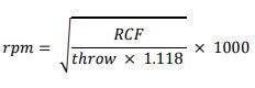
Where:
rpm = shaker speed (revolutions per minute)
RCF = relative centrifugal force (g), provided in Table 2 for various cultureware
throw = shaking diameter (mm), as specified by manufacturer - Perform a full-medium change every 3 - 4 days as follows:
a.Tilt the cultureware.
b.Using a 5 mL serological pipette at the slowest setting, slowly remove medium.
c.Add 3 mL/well of fresh Maturation Medium.
d.Return plate to the orbital shaker in a 37°C incubator. - Cerebral organoids will continue to grow and reach a size of approximately 3 - 4 mm in diameter after 30 days in Maturation Medium.
- Day 10: Organoids display budding morphology, which indicates expanding neuropithelia.
- Day 15: The expanded neuroepithelial buds merge and the organoid displays a denser core.
- Day 20: Organoids will be over 750 μm in diameter; small rosettes may be observed.
- Day 30: Organoids will be > 1 mm in diameter and will have a dense core with the appearance of layered structures.
- Day 40: Organoids will be approximately 3 - 5 mm in diameter. At this point the organoids are very dense and dark in the center and maintain cortical layering. Organoids that do not form properly display clear cysts and do not reach a size of > 1 mm by day 40. This is the typical timepoint where we observe cortical layering with PAX-6+ progenitors lining a central ventricle distinctly separated from TUJ-1+ neurons.
- Cerebral organoids can continue to be cultured beyond day 40 using STEMdiff™ Cerebral Organoid Maturation Kit (Catalog #08571).
References
- Lancaster et al. (2013) Cerebral organoids model human brain development and microcephaly. Nature 501(7467): 373–9.
- Lancaster et al. (2014) Organogenesis in a dish: modeling development and disease using organoid technologies. Science 345(6194): 1247125.
Request Pricing
Thank you for your interest in this product. Please provide us with your contact information and your local representative will contact you with a customized quote. Where appropriate, they can also assist you with a(n):
Estimated delivery time for your area
Product sample or exclusive offer
In-lab demonstration


