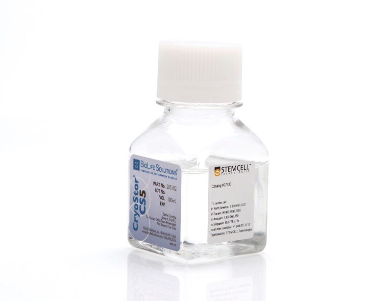
CryoStor® CS5

Animal component-free, defined cryopreservation medium with 5% DMSO

CryoStor® CS5

Animal component-free, defined cryopreservation medium with 5% DMSO
From: 380 USD
Catalog #
(Select a product)
Animal component-free, defined cryopreservation medium with 5% DMSO
Add to Wish List

Product Advantages


  • Ready-to-use

  • Serum-free and protein-free

  • Animal component-free

  • cGMP manufactured with USP grade/highest-quality components

  • FDA master file

  • Sterility, endotoxin, and cell-based quality control testing

Overview

Maximize post-thaw cell recovery and viability following cryopreservation at low temperatures (-80°C to -196°C) with ready-to-use CryoStor® CS5. Serum-free, animal component-free, and cGMP-manufactured, this defined medium provides a safe, protective environment for cells and tissues during freezing and thawing processes and storage. Available in a variety of convenient formats, CryoStor® CS5 is formulated with USP-grade components and contains 5% DMSO.
Contains
• 5% dimethyl sulfoxide (DMSO)
• Other ingredients
Cell Type
CHO Cells, Mesenchymal Stem and Progenitor Cells, Other, Pluripotent Stem Cells
Species
Human, Mouse, Non-Human Primate, Other, Rat
Application
Cryopreservation
Brand
CryoStor
Area of Interest
Immunology, Stem Cell Biology
Formulation Category
Animal Component-Free, Serum-Free

Protocols and Documentation

Find supporting information and directions for use in the Product Information Sheet or explore additional protocols below.

Document Type
Product Name
Catalog #
Lot #
Language
Product Name
CryoStor® CS5
Catalog #
07953, 07933, 07949
Lot #
All
Language
English
Document Type
Safety Data Sheet
Product Name
CryoStor® CS5
Catalog #
07953, 07933, 07949
Lot #
All
Language
English

Resources and Publications

Publications (11)

Single-cell vector copy number analysis of phenotypically defined long-term hematopoietic stem cells for gene therapy safety assessment. Y. Sato et al. Molecular therapy. Advances 2026 Sep

Abstract

Hematopoietic stem cell (HSC)-based gene therapy has emerged as a transformative approach for the treatment of genetic diseases; however, accurate evaluation of vector copy number (VCN) remains critical for ensuring safety. Conventional bulk VCN assays, including quantitative PCR (qPCR) and droplet digital PCR (ddPCR), do not resolve clonal heterogeneity and cannot identify rare high-VCN cells that may contribute disproportionately to insertional mutagenesis risk. Here, we developed an accessible single-cell VCN profiling method by combining fluorescence-activated cell sorting (FACS) of phenotypically defined long-term HSCs (Lineage- CD34+ CD38- CD90+ CD45RA- cells) with whole-genome amplification followed by conventional qPCR. This approach enabled resolution of VCN distributions at single-cell level using standard laboratory techniques. Notably, single-cell analysis revealed a high VCN tail that bulk VCN analysis could not resolve. Furthermore, in a humanized mouse transplantation model, single-cell VCN profiling demonstrated that overall VCN distributions could be analyzed after engraftment, although inter-donor and inter-mouse variability was observed. Collectively, this method provides a rapid, cost-effective, and phenotypically resolved strategy for assessing VCN heterogeneity in gene-modified HSCs. Single-cell VCN profiling offers complementary insights beyond conventional bulk assays and may enhance preclinical safety evaluation of gene and cell therapy products.
CD105+ fibroblasts support an immunosuppressive niche in women at high risk of breast cancer initiation E. Carlson et al. Breast Cancer Research : BCR 2025 May

Abstract

BackgroundAging is the greatest risk factor for breast cancer, and although epithelial cells are the source of carcinomas, epithelial changes alone do not fully explain cancer susceptibility. Fibroblasts and macrophages are key stromal constituents around the cells of origin for cancer in breast tissue. With age, macrophages surrounding terminal ductal lobular units (TDLUs) become increasingly immunosuppressive. CD105+ fibroblasts intercalate within TDLUs, drive luminal differentiation, and give rise to immunosuppressive cancer-associated fibroblasts in other tissues. We propose that differences in fibroblasts are a crucial component of the stroma that shapes cancer susceptibility.MethodsPrimary peri-epithelial fibroblast cultures were established from prophylactic and reduction mammoplasties from 30 women ranging in age from 16 to 70 years and from BRCA1 mutation carriers. Growth characteristics, transcriptional profiles, differentiation potential, and secreted proteins were profiled for fibroblast subtypes from diverse donors. Co-cultures with fibroblasts, macrophages, and T cells were used to ascertain the functional role played by CD105+ fibroblasts in immune cell modulation.ResultsWe found that peri-epithelial CD105+ fibroblasts are enriched in older women as well as women who carry BRCA1 mutations. These CD105+ fibroblasts exhibit robust adipogenesis and secrete factors related to macrophage polarization. Macrophages cocultured with fibroblasts better maintain or enhance polarization states than media alone. CD105+ fibroblasts increased expression of immunosuppressive macrophage genes. CD105+ fibroblasts supported anti-inflammatory macrophage-mediated suppression of T cell proliferation, whereas CD105− fibroblasts significantly reduced the suppressive effect of anti-inflammatory macrophages on T cell proliferation.ConclusionsEstablishment of a coculture system to dissect the molecular circuits between CD105+ fibroblasts and macrophages that drive immunosuppressive macrophage polarization has broad utility in understanding mammary gland development and events that precede cancer initiation. CD105+ fibroblasts and macrophages may coordinate to suppress immunosurveillance and increase breast cancer susceptibility.Supplementary InformationThe online version contains supplementary material available at 10.1186/s13058-025-02040-7.
Inflammatory Responses and Barrier Function of Endothelial Cells Derived from Human Induced Pluripotent Stem Cells. O. V. Halaidych et al. Stem cell reports 2018 MAY

Abstract

Several studies have reported endothelial cell (EC) derivation from human induced pluripotent stem cells (hiPSCs). However, few have explored their functional properties in depth with respect to line-to-line and batch-to-batch variability and how they relate to primary ECs. We therefore carried out accurate characterization of hiPSC-derived ECs (hiPSC-ECs) from multiple (non-integrating) hiPSC lines and compared them with primary ECs in various functional assays, which included barrier function using real-time impedance spectroscopy with an integrated assay of electric wound healing, endothelia-leukocyte interaction under physiological flow to mimic inflammation and angiogenic responses in in vitro and in vivo assays. Overall, we found many similarities but also some important differences between hiPSC-derived and primary ECs. Assessment of vasculogenic responses in vivo showed little difference between primary ECs and hiPSC-ECs with regard to functional blood vessel formation, which may be important in future regenerative medicine applications requiring vascularization.